Brain activity has an unmistakable signature: the firing of neurons, as brain cells relay information to one another through the triggered release of chemical neurotransmitters, which are received by the long, branching dendrites of neighbouring cells.
This microscopic ritual is distinctive, but it doesn’t belong only to neurons, scientists have now found. Researchers have discovered a previously unnoticed, similar-looking signalling process taking place outside the nervous system, observing bursts of neuron-like activity in certain skin cells.
The team from Rockefeller University observed these interactions between two different kinds of skin cells: melanocytes, which produce the UV-absorbing pigment melanin; and keratinocytes, which make up the vast majority of the epidermis, protecting the body from environmental exposure, partially via melanin.
“Keratinocytes are known regulators of melanocyte behaviour, and much work has been done to understand how keratinocytes influence melanocyte cell proliferation and the production and transfer of pigment throughout the skin,” the authors write in their new study.
“Nonetheless, cell-cell communication between melanocytes and keratinocytes, at the single-cell level, is poorly understood.”
In experiments with the two types of skin cells grown together in co-cultures – and also investigations of samples of intact human skin – the researchers have gotten us closer to seeing how this process actually works, and the surprise is that it appears to resemble neural communication.
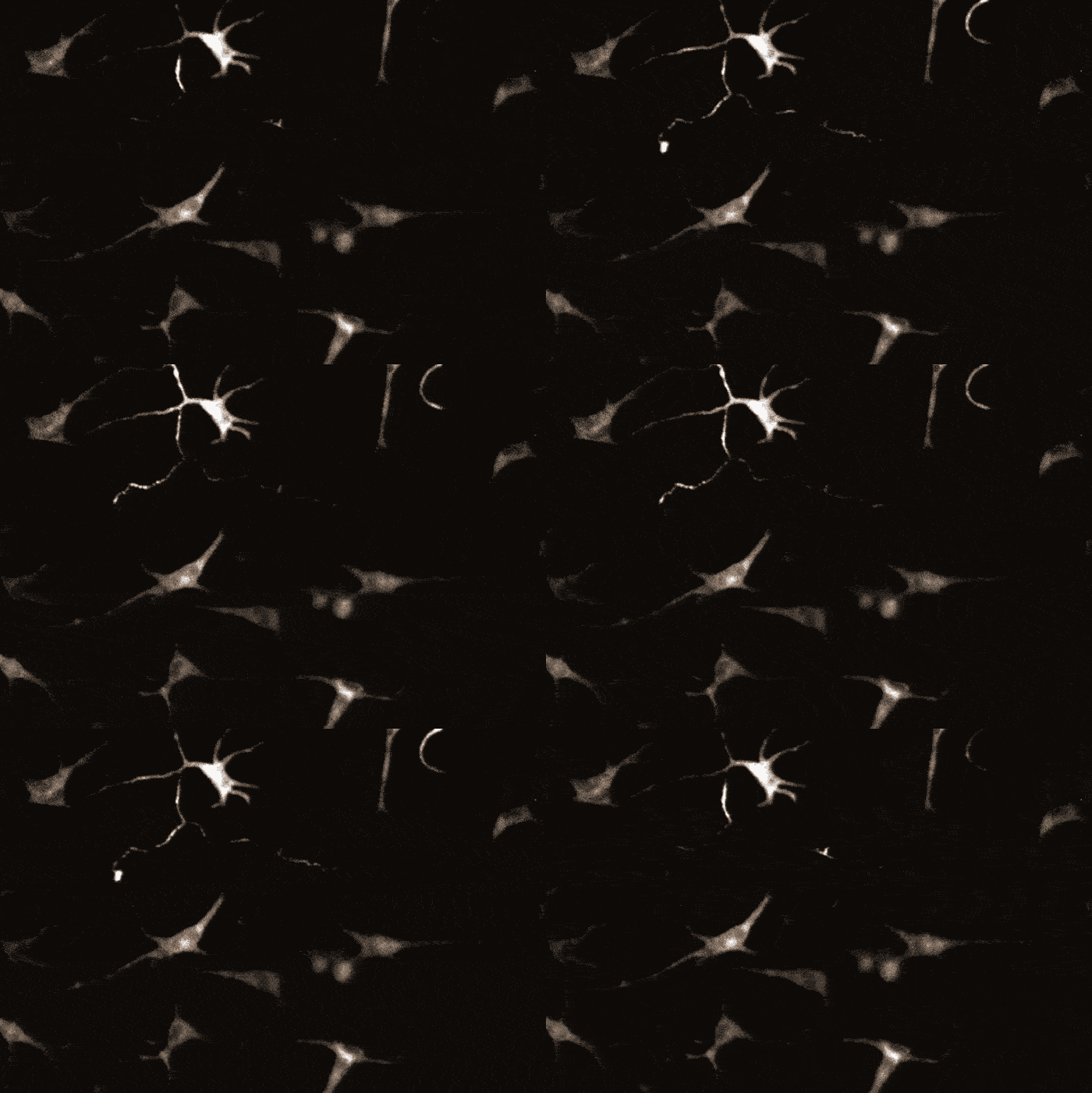
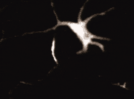
 Calcium signalling in skin cells. (Belote and Simon, Journal of Cell Biology, 2019)
“We saw that keratinocytes wrap around melanocytes, forming intimate connections that reminded us of neurons,” says cellular biophysicist Sanford M. Simon.
In the study, the researchers observed that chemical signals from keratinocytes trigger signals called calcium transients within the melanocyte dendrites.
This calcium signalling process, sparked by the production of two keratinocyte secretions – endothelin and acetylcholine – was also observed in smaller dendritic spine-like structures on the melanocytes, which the researchers say can also be seen in intact human skin.
Other kinds of cells have exhibited signalling systems that resemble this before, but it wasn’t known that skin cells could do it, as it’s primarily associated with neuronal functioning.
“This type of localised cell-cell communication has been viewed as a hallmark of the nervous system,” the researchers explain.
“Dendritic morphologies are not unique to the nervous system, but it is not known if non-neuronal dendrites, such as those on melanocytes, are also capable of compartmentalising signals received from adjacent cells.”
According to this new evidence, they are. Together with the new discovery of the spine-like structures protruding from the dendrites – which the team says are “strikingly similar” to neuronal dendritic spines – the findings suggest the existence of a deeper complexity in skin cell communication that scientists never knew about.
“There’s a very sophisticated level of signalling going on here that we did not appreciate,” says Simon.
“This opens up a lot of exciting questions about the basic physiology of the skin.”
[“source=sciencealert”]
